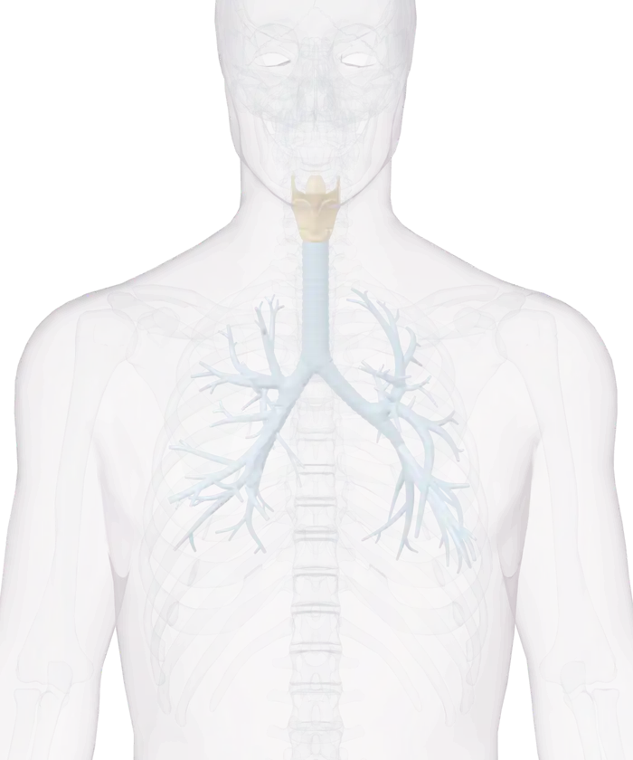
28 cartilages, ligaments and airways. A part that comes as a left and right pair is listed once.
Smaller but thicker and stronger than the thyroid cartilage, the cricoid cartilage forms the lower and back parts of the wall of the larynx. It has a narrow arch in front and a deep, broad plate (lamina) behind. Its lower border is joined to the first ring of the trachea by the cricotracheal ligament.
Segmental bronchi are branches of the bronchial tree inside the lungs. The trachea divides into the right and left primary bronchi, which branch into smaller and smaller airways ending in tiny air sacs called alveoli. As the branching continues, the walls hold less cartilage and more smooth muscle.
Nearly 5 cm long, the left main bronchus is narrower but longer than the right. It passes beneath the arch of the aorta and crosses in front of the esophagus, the thoracic duct and the descending aorta. The bronchus enters the root of the left lung level with the sixth thoracic vertebra.
Wider, shorter and more upright than the left, the right main bronchus is about 2.5 cm long. It enters the right lung nearly level with the fifth thoracic vertebra. About 2 cm from its start it gives a branch to the upper lobe, then divides into branches for the middle and lower lobes.
Commonly called the windpipe, the trachea is the main airway to the lungs. At the level of the fifth thoracic vertebra it divides into the right and left bronchi, and the esophagus lies directly behind it. Cartilage in its wall keeps the trachea from collapsing, while mucus in its lining traps airborne particles and cilia sweep the mucus upward.
At the back of the tongue sits the epiglottis, a stiff yet flexible piece of cartilage covered with a lining of tissue. During swallowing it closes over the windpipe (trachea) so that food does not enter the airway. This closing helps prevent coughing or choking after a swallow.
An elastic band, the hyo-epiglottic ligament connects the epiglottis with the hyoid bone. It runs from the lower part of the front surface of the epiglottis to the upper border of the body of the hyoid bone. This band belongs to the extrinsic ligaments of the larynx, which link its cartilages to the hyoid bone and the trachea.
The cuneiform cartilages are two small, elongated pieces of yellow elastic cartilage, one on each side of the larynx. Each lies in the aryepiglottic fold, a fold of mucous membrane stretched between the side of the epiglottis and the tip of the arytenoid cartilage. There it raises a small whitish bump on the surface, just in front of the arytenoid cartilage.
Two arytenoid cartilages sit at the back of the larynx, on the upper border of the plate of the cricoid cartilage. Each is shaped like a pyramid. The front corner of its base, the vocal process, anchors the vocal ligament, and the outer corner, the muscular process, receives two cricoarytenoid muscles.
Two small, cone-shaped nodules of yellow elastic cartilage, the corniculate cartilages sit on the tips of the arytenoid cartilages at the back of the larynx. They extend the arytenoid cartilages backward and toward the midline. Each lies in the back part of an aryepiglottic fold of mucous membrane and is sometimes fused with the arytenoid cartilage.
Made mainly of yellow elastic tissue, the conus elasticus (cricothyroid membrane) links the thyroid, cricoid and arytenoid cartilages. Its thin side portions lie just under the lining of the larynx. Each runs from the upper border of the cricoid cartilage to the vocal ligament, which forms its free edge.
A broad sheet of fibrous and elastic tissue, the thyrohyoid membrane joins the upper border of the thyroid cartilage to the hyoid bone. Its thicker middle part is the median thyrohyoid ligament. A round elastic cord, the lateral thyrohyoid ligament, forms its back border on each side and reaches the greater horn of the hyoid bone.
Inside each vocal fold (true vocal cord) runs a vocal ligament, a strong band of yellow elastic tissue. It stretches from the angle of the thyroid cartilage in front to the vocal process of the arytenoid cartilage behind. The vocal folds, which enclose the two ligaments, take part in producing sound.
Thick and strong, the median cricothyroid ligament is the front part of the conus elasticus (cricothyroid membrane). Narrow at the top and broad at the bottom, it joins the front of the thyroid cartilage to the front of the cricoid cartilage. A cricothyroid muscle overlaps it on each side, and between the two muscles it lies just under the skin.
The stem of the epiglottis is fastened to the thyroid cartilage by the thyro-epiglottic ligament, a long, slender elastic cord. The cord attaches at the angle where the two plates of the thyroid cartilage meet, just beneath the superior thyroid notch. The epiglottis itself is a thin, leaf-shaped plate of fibrocartilage in front of the entrance to the larynx.
Largest of the cartilages of the larynx, the thyroid cartilage is made of two plates (laminae). The plates join at an angle in the midline, forming a bump under the skin: the laryngeal prominence, or Adam's apple. The bump is larger in males, and membranes and ligaments tie the cartilage to the hyoid bone and the cricoid cartilage.