Skip to the main content
3D Body Visualizer

3D eye model: each eye in 21 parts, with the outer ear

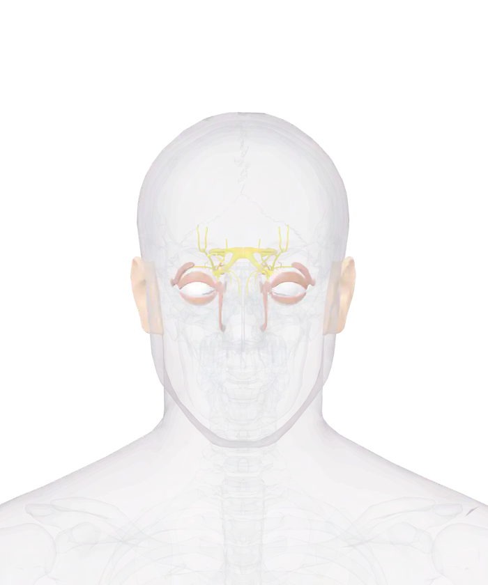
The eyes of the 3D anatomy model inside a glass skull, seen from the front

Pick a part

Tap or click a part of the eye for its name and its size. Hide the eyelids and the sclera to reach the lens, the iris and the retina, or pull a part out and turn it around.

Eye and ear: 43 parts

Switch a group on or off, or open its list to pick a part by name.

  • Groups in the eye and ear
  • Groups in the nervous system
See the full human anatomy

The eyes are those of one adult man, from the BodyParts3D project. Of the ear the model has only the outer ears: the eardrum, the three small bones and the inner ear are missing.

The eye and ear group of the model has 43 parts: 20 in the 2 eyeballs, 22 in the eyelids and tear system, and the outer ears. Each sclera, the white coat of the eye, is 2.6 cm across. The view also shows 37 nerves of the eye socket, among them both optic nerves.

The eye and ear of the model in 4 groups

Each eye of the model has 10 parts in the eyeball and 11 in the eyelids and tear system.
Group Parts What it holds
Eyeball 20 For each eye: cornea, iris, lens, retina, choroid, sclera, vitreous body and 3 more
Eyelids and tear system 22 For each eye: the plates of both eyelids, the tear gland, the tear ducts and 6 more
Outer ear 1 Both outer ears as one part
Nerves of the eye socket 37 The optic nerves and the nerves of the eye muscles

The skull is drawn as glass, so each eyeball shows in its socket. To look inside an eye, tap the sclera and press Hide it, then do the same with the choroid.

How the eye works

Light passes through 4 parts of the eye on its way to the brain, according to the National Eye Institute.

Light passes the cornea, the pupil and the lens before it reaches the retina.
Part What it does In the model
Cornea The clear dome at the front, which bends light 1.4 cm across
Pupil and iris The iris controls how much light enters through the pupil Iris, 1.3 cm across
Lens Works with the cornea to focus light on the retina 1.0 cm across
Retina Light-sensitive tissue that turns light into electrical signals Lines the back of each eyeball

The optic nerve then carries the signals to the brain. It is a bundle of more than 1 million nerve fibers. The macula is a small area of the retina needed for central vision, and the vitreous is the clear gel that fills the eye.

Six muscles move each eyeball: 4 rectus muscles and 2 oblique muscles. The model lists them with the muscular system, so they show in the 3D muscular system view and in the full 3D anatomy.

How the ear works

Hearing turns sound waves into electrical signals in a series of steps, according to the National Institute on Deafness and Other Communication Disorders.

Sound passes 3 parts of the ear, and the model draws the first.
Part of the ear What happens there In the model
Outer ear Sound travels through the ear canal to the eardrum Yes, the outer ears
Middle ear The eardrum passes vibrations to 3 small bones No
Inner ear The cochlea turns the vibrations into electrical signals No

The 3 bones are the malleus, the incus and the stapes. The cochlea is a fluid-filled structure shaped like a snail. Hair cells along its length detect different pitches, and the auditory nerve carries their signals to the brain.

What the model leaves out

Of the ear, the model has the outer ears only. The eardrum, the 3 small bones and the inner ear are missing. The eyes are those of one adult man.

The brain that the optic nerves run to is in the 3D nervous system. The 6 muscles of each eye are in the 3D muscular system, and the skin of the eyelids is part of the 3D skin view.

Every part of the eye and ear in the model by name

41 parts of the eye, its nerves and the outer ear. A part that comes as a left and right pair is listed once.

Nervous system: nerves of the eye socket (19)
  • Anterior ethmoidal nerve
  • Ciliary ganglion
  • Communicating branch of nasociliary nerve with ciliary ganglion
  • Frontal nerve
  • Inferior branch of oculomotor nerve
  • Infratrochlear nerve
  • Lacrimal nerve
  • Long ciliary nerve
  • Nasociliary nerve
  • Ophthalmic nerve
  • Optic chiasm
  • Optic nerve
  • Optic tract
  • Posterior ethmoidal nerve
  • Short ciliary nerve
  • Superior branch of oculomotor nerve
  • Supra-orbital nerve
  • Supratrochlear nerve
  • Trochlear nerve
Eye and ear: eyeball (10)
  • Anterior chamber of eyeball
  • Choroid
  • Cornea
  • Corona ciliaris
  • Iris
  • Lens
  • Optic part of retina
  • Sclera
  • Suspensory ligament of lens
  • Vitreous body
Eye and ear: eyelids and tear system (11)
  • Check ligament of lateral rectus
  • Check ligament of medial rectus
  • Common tendinous ring
  • Lacrimal canaliculus
  • Lacrimal gland
  • Lacrimal lake
  • Lacrimal sac
  • Nasolacrimal duct
  • Tarsal plate of lower eyelid
  • Tarsal plate of upper eyelid
  • Trochlea of superior oblique
Eye and ear: outer ear (1)
  • Outer ears

More to explore

Questions people ask

How does light travel through the eye?

Light enters the eye through the cornea, passes through the pupil, which is the opening at the center of the iris, and is focused by the lens onto the retina. The retina turns the light into electrical signals, and the optic nerve carries the signals to the brain.

How many nerve fibers are in the optic nerve?

The optic nerve is a bundle of more than 1 million nerve fibers, according to the National Eye Institute. The model draws the left and the right optic nerve, the crossing where they meet and the 2 optic tracts behind it.

How many muscles move the eye?

Six muscles move each eyeball: the medial, lateral, superior and inferior rectus and the superior and inferior oblique. The model lists the 6 muscles of each eye with the muscular system, in the 3D muscular system view.

What are the three bones of the middle ear?

The three bones of the middle ear are the malleus, the incus and the stapes. They pass the vibrations of the eardrum on to the cochlea. The model has the outer ears only and none of the 3 bones.

What is the white of the eye called?

The white of the eye is the sclera, the white outer coat of the eyeball. Each sclera of the model is 2.6 cm across. Hide the sclera to see the choroid, the retina and the vitreous body inside it.

5 sources
  1. How the Eyes Work, National Eye Institute. Accessed 14 November 2026.
  2. Glossary, National Eye Institute. Accessed 14 November 2026.
  3. Anatomy, Head and Neck: Eye Muscles, StatPearls Publishing, on the NCBI Bookshelf of the National Library of Medicine. Accessed 14 November 2026.
  4. How Do We Hear?, National Institute on Deafness and Other Communication Disorders. Accessed 14 November 2026.
  5. BodyParts3D, Database Center for Life Science (DBCLS), Japan. Accessed 14 November 2026.