Skip to the main content
3D Body Visualizer

3D heart model and circulatory system: 656 named parts

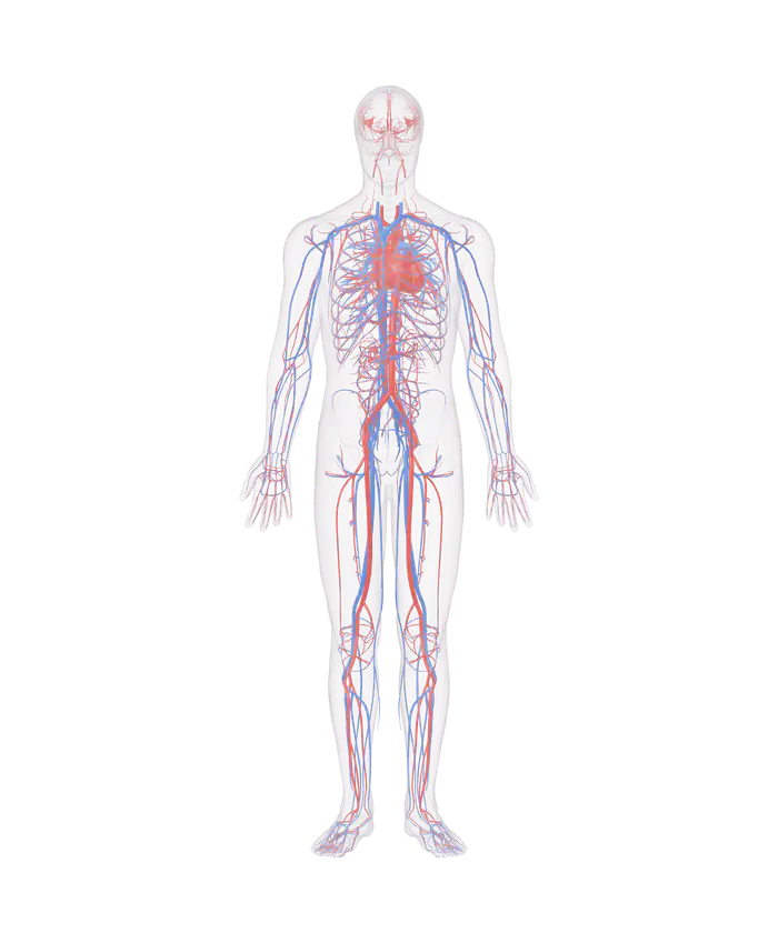
The heart, arteries and veins of the 3D anatomy model inside a glass skeleton, seen from the front

Pick a part

Tap or click the heart, an artery or a vein for its name and its size. Fade the rest to follow one vessel, or hide the veins to see the arteries alone.

Cardiovascular system: 656 parts

Switch a group on or off, or open its list to pick a part by name.

  • Groups in the cardiovascular system
See the full human anatomy

The heart and vessels are those of one adult man, from the BodyParts3D project. The smallest vessels and the capillaries are not in the model, and the course of vessels differs from person to person.

The cardiovascular system of the model has 656 parts: a heart in 23 parts, 26 coronary vessels, 369 arteries, 177 veins and 61 pulmonary vessels. The longest single vessel is the great saphenous vein, which runs 82.2 cm up each leg of this body. Tap any part for its name and its size.

The cardiovascular system in 6 groups

The model has 369 arteries and 177 veins, and the heart alone is drawn in 23 parts.
Group Parts What it holds
Heart 23 The 4 chambers, the walls, the valve leaflets and the papillary muscles
Coronary vessels 26 The arteries and veins of the heart's own wall
Arteries 369 The aorta and its branches to the head, trunk, arms and legs
Veins 177 The veins that return blood to the heart, among them the saphenous and cephalic veins
Pulmonary arteries 33 The vessels from the right ventricle into both lungs
Pulmonary veins 28 The vessels from the lungs back to the left atrium

Arteries are drawn red and veins blue. The pulmonary vessels reverse the colors: the pulmonary arteries are blue and the pulmonary veins red. Each of the 6 groups has its own switch beside the model, so the veins can be hidden to leave the arteries alone.

How the heart is built

The heart is a muscular pump with 4 chambers, and the National Cancer Institute gives its size and shape as those of a closed fist. Its wall has 3 layers: the epicardium on the outside, the myocardium, which is the muscle, and the endocardium on the inside. About two-thirds of its mass lies to the left of the midline.

The four valves of the heart are drawn as 11 separate leaflets and cusps.
Valve Where it sits In the model
Tricuspid valve Between the right atrium and the right ventricle 3 leaflets: anterior, posterior and septal
Mitral valve Between the left atrium and the left ventricle 2 leaflets: anterior and posterior
Pulmonary valve Between the right ventricle and the pulmonary trunk 3 cusps
Aortic valve Between the left ventricle and the aorta 3 cusps

The heart sets its own pace. The sinoatrial node starts an impulse 70 to 80 times a minute without any signal from a nerve, and one cardiac cycle lasts 0.8 second at a normal heart rate. Hide the walls of the ventricles in the model to see the valve leaflets and the papillary muscles inside.

Arteries, capillaries and veins

There are 3 types of blood vessel. Arteries carry blood away from the heart, capillaries exchange nutrients and waste with the tissue, and veins carry blood back to the heart. The aorta has 3 portions: the ascending aorta, the arch and the descending aorta. The descending aorta of the model is 33.5 cm long.

Blood travels in 2 circuits. The pulmonary circuit runs from the right ventricle to the lungs and back to the left atrium. The systemic circuit runs from the left ventricle to the rest of the body and back to the right atrium.

To follow one vessel, type its name into the search box, pick it and press Fade the rest. The vessel stays solid while the body turns to glass around it.

What the model leaves out

The model has no capillaries, which the National Cancer Institute calls the smallest and most numerous of the vessels. Many of the smaller arteries and veins carry a name and a size only, with no written description. The vessels are those of one adult man, and their course differs from person to person.

The 3D anatomy view shows the heart together with the other organs, and the 3D respiratory system shows the airways that the pulmonary vessels follow into the lungs. The spleen, which filters blood, is in the 3D lymphatic system.

Every part of the heart and every vessel in the model by name

410 heart parts, arteries and veins. A part that comes as a left and right pair is listed once.

Heart (18)
  • Anterior cusp of aortic valve
  • Anterior cusp of pulmonary valve
  • Anterior leaflet of mitral valve
  • Anterior leaflet of tricuspid valve
  • Anterior papillary muscle of ventricle
  • Anterolateral head of lateral papillary muscle of ventricle
  • Cavity of atrium
  • Cavity of ventricle
  • Lateral papillary muscle of ventricle
  • Posterior cusp of aortic valve
  • Posterior cusp of pulmonary valve
  • Posterior leaflet of mitral valve
  • Posterior leaflet of tricuspid valve
  • Posterior papillary muscle of ventricle
  • Septal leaflet of tricuspid valve
  • Septal papillary muscle of ventricle
  • Wall of atrium
  • Walls of the ventricles
Coronary vessels (24)
  • Anterior cardiac vein
  • Anterior interventricular vein
  • Circumflex branch of coronary artery
  • Conus artery
  • Conus branch of anterior interventricular branch of coronary artery
  • Coronary sinus
  • Diagonal branch of anterior descending branch of coronary artery
  • First anterior branch of anterior interventricular branch of coronary artery
  • First anterior ventricular branch of coronary artery
  • First posterior ventricular branch of coronary artery
  • First septal branch of posterior interventricular artery
  • Great cardiac vein
  • Marginal branch of coronary artery
  • Marginal vein
  • Middle cardiac vein
  • Posterior interventricular branch of coronary artery
  • Posterior vein of ventricle
  • Second anterior branch of anterior interventricular branch of coronary artery
  • Second septal branch of posterior interventricular artery
  • Septal branch of anterior interventricular artery
  • Small cardiac vein
  • Third anterior branch of anterior interventricular branch of coronary artery
  • Trunk of anterior interventricular branch of coronary artery
  • Trunk of coronary artery
Arteries (209)
  • Acromial branch of thoraco-acromial artery
  • Anterior cecal artery
  • Anterior cerebral artery
  • Anterior choroidal artery
  • Anterior circumflex humeral artery
  • Anterior communicating artery
  • Anterior division of renal artery
  • Anterior inferior cerebellar artery
  • Anterior inferior pancreaticoduodenal artery
  • Anterior inferior segmental hepatic artery
  • Anterior interosseous artery
  • Anterior parietal artery
  • Anterior spinal artery
  • Anterior superior pancreaticoduodenal artery
  • Anterior superior segmental hepatic artery
  • Anterior temporal artery
  • Anterior temporal branch of lateral occipital artery
  • Anterior tibial artery
  • Anterior tibial recurrent artery
  • Anterior ulnar recurrent artery
  • Anterolateral central branch of middle cerebral artery
  • Appendicular artery
  • Arch of aorta
  • Arcuate artery
  • Arteria princeps pollicis
  • Arteria radialis indicis
  • Artery of central sulcus
  • Artery of postcentral sulcus
  • Artery of precentral sulcus
  • Ascending aorta
  • Ascending branch of colic artery
  • Ascending branch of inferior branch of ileocolic artery
  • Axillary artery
  • Basilar artery
  • Brachial artery
  • Brachiocephalic artery
  • Branch of anterior choroidal artery to posterior limb of internal capsule
  • Branch of lateral circumflex femoral artery
  • Branch of middle cerebral artery to angular gyrus
  • Bronchial artery
  • Bronchial branch of arch of aorta
  • Callosomarginal artery
  • Caudal pancreatic artery
  • Caudate lobe branch of hepatic artery
  • Celiac trunk
  • Circumflex scapular artery
  • Colic artery
  • Common carotid artery
  • Common hepatic artery
  • Common iliac artery
  • Common interosseous artery
  • Common plantar digital arteries
  • Costocervical trunk
  • Deep brachial artery
  • Deep cervical artery
  • Deep palmar arch
  • Deep plantar artery
  • Deltoid branch of thoraco-acromial artery
  • Descending aorta
  • Descending branch of colic artery
  • Descending branch of lateral circumflex femoral artery
  • Descending genicular artery
  • Distal perforating artery
  • Dorsal carpal branch of radial artery
  • Dorsal carpal branch of ulnar artery
  • Dorsal digital arteries of hand
  • Dorsal digital artery of foot
  • Dorsal metacarpal arteries
  • Dorsal pancreatic artery
  • Dorsal scapular artery
  • Dorsalis pedis artery
  • Esophageal artery
  • Esophageal branches of thoracic aorta
  • External iliac artery
  • Femoral artery
  • Fibular artery
  • First common palmar digital artery
  • First posterior intercostal artery
  • Fourth common palmar digital artery
  • Gastric artery
  • Gastro-epiploic artery
  • Great pancreatic artery
  • Hepatic artery proper
  • Hypothalamic branch of posterior communicating artery
  • Ileal branch of inferior branch of ileocolic artery
  • Ileocolic artery
  • Inferior epigastric artery
  • Inferior lateral genicular artery
  • Inferior medial genicular artery
  • Inferior mesenteric artery
  • Inferior pancreatic artery
  • Inferior pancreaticoduodenal artery
  • Inferior phrenic artery
  • Inferior suprarenal artery
  • Inferior thyroid artery
  • Inferior ulnar collateral artery
  • Insular part of middle cerebral artery
  • Intermediomedial branch of callosomarginal artery
  • Internal carotid artery
  • Internal iliac artery
  • Internal thoracic artery
  • Lateral branch of pontine artery
  • Lateral branch of superior cerebellar artery
  • Lateral circumflex femoral artery
  • Lateral frontobasal artery
  • Lateral inferior segmental hepatic artery
  • Lateral occipital artery
  • Lateral plantar artery
  • Lateral proper palmar digital artery of little finger
  • Lateral proper palmar digital artery of middle finger
  • Lateral proper palmar digital artery of ring finger
  • Lateral superior genicular artery
  • Lateral superior segmental hepatic artery
  • Lateral tarsal artery
  • Lateral thoracic artery
  • Lumbar artery
  • Marginal artery of colon
  • Medial branch of pontine artery
  • Medial branch of superior cerebellar artery
  • Medial frontobasal artery
  • Medial inferior segmental hepatic artery
  • Medial occipital artery
  • Medial plantar artery
  • Medial proper palmar digital artery of index finger
  • Medial proper palmar digital artery of middle finger
  • Medial proper palmar digital artery of ring finger
  • Medial superior genicular artery
  • Medial superior segmental hepatic artery
  • Middle colic artery
  • Middle collateral branch of deep brachial artery
  • Middle genicular artery
  • Middle suprarenal artery
  • Middle temporal branch of lateral occipital artery
  • Middle temporal branch of middle cerebral artery
  • Musculophrenic artery
  • Ophthalmic artery
  • Palmar metacarpal artery
  • Paracentral branch of callosomarginal artery
  • Pectoral branch of thoraco-acromial artery
  • Perforating arteries of thigh
  • Pericallosal artery
  • Plantar arch
  • Plantar metatarsal artery
  • Polar temporal artery
  • Popliteal artery
  • Postcommunicating part of posterior cerebral artery
  • Posterior cecal artery
  • Posterior circumflex humeral artery
  • Posterior communicating artery
  • Posterior division of renal artery
  • Posterior inferior cerebellar artery
  • Posterior inferior pancreaticoduodenal artery
  • Posterior inferior segmental hepatic artery
  • Posterior intercostal arteries
  • Posterior medial choroidal artery
  • Posterior parietal artery
  • Posterior superior pancreaticoduodenal artery
  • Posterior superior segmental hepatic artery
  • Posterior temporal branch of middle cerebral artery
  • Posterior temporal branches of lateral occipital artery
  • Posterior tibial artery
  • Posterior ulnar recurrent artery
  • Posteromedial branch of callosomarginal artery
  • Posteromedial central branch of posterior cerebral artery
  • Precommunicating part of posterior cerebral artery
  • Precuneal branch of pericallosal artery
  • Prefrontal artery
  • Proper plantar digital arteries of foot
  • Radial artery
  • Radial collateral branch of deep brachial artery
  • Radial recurrent artery
  • Recurrent interosseous artery
  • Renal artery
  • Second common palmar digital artery
  • Second posterior intercostal artery
  • Sigmoid artery
  • Sphenoid part of middle cerebral artery
  • Splenial artery
  • Splenic artery
  • Subclavian artery
  • Subcostal artery
  • Subscapular artery
  • Superficial cervical artery
  • Superficial epigastric artery
  • Superficial medial plantar artery
  • Superficial palmar arterial arch
  • Superior cerebellar artery
  • Superior epigastric artery
  • Superior intercostal artery
  • Superior mesenteric artery
  • Superior rectal artery
  • Superior ulnar collateral artery
  • Superior vermian branch of medial branch of superior cerebellar artery
  • Suprascapular artery
  • Temporo-occipital branch of middle cerebral artery
  • Testicular artery
  • Thalamogeniculate artery
  • Thalamoperforating artery
  • Third common palmar digital artery
  • Thoracodorsal artery
  • Thyrocervical trunk
  • Transverse cervical artery
  • Trunk of gastroduodenal artery
  • Trunk of hepatic artery
  • Trunk of inferior terminal branch of middle cerebral artery
  • Trunk of thoraco-acromial artery
  • Ulnar artery
  • Ureteric segment of renal artery
  • Vertebral artery
Veins (106)
  • Accessory hemiazygos vein
  • Anterior circumflex humeral vein
  • Anterior inferior segmental branch of portal vein
  • Anterior inferior segmental tributary of middle hepatic vein
  • Anterior intercostal veins
  • Anterior superior segmental branch of portal vein
  • Anterior superior segmental tributary of hepatic vein
  • Anterior tibial vein
  • Ascending lumbar vein
  • Axillary vein
  • Azygos vein
  • Basilic vein
  • Brachiocephalic vein
  • Caudate lobe branch of portal vein
  • Cephalic vein
  • Circumflex scapular vein
  • Colic vein
  • Common iliac vein
  • Common palmar digital vein
  • Deep femoral vein
  • Deep palmar venous arch
  • Dorsal digital veins
  • Dorsal metatarsal veins of foot
  • Dorsal venous arch of foot
  • Dorsal venous network of hand
  • External iliac vein
  • Femoral vein
  • Fibular vein
  • Gastric vein
  • Gastroepiploic vein
  • Genicular vein
  • Great saphenous vein
  • Hemiazygos vein
  • Hepatic portal vein
  • Ileal vein
  • Ileocolic vein
  • Iliolumbar vein
  • Inferior epigastric vein
  • Inferior gluteal vein
  • Inferior mesenteric vein
  • Inferior phrenic vein
  • Inferior vena cava
  • Internal iliac vein
  • Internal jugular vein
  • Internal pudendal vein
  • Internal thoracic vein
  • Lateral circumflex femoral vein
  • Lateral inferior segmental branch of portal vein
  • Lateral inferior segmental tributary of hepatic vein
  • Lateral sacral vein
  • Lateral superior segmental branch of portal vein
  • Lateral superior segmental tributary of hepatic vein
  • Lateral thoracic vein
  • Lumbar vein
  • Medial brachial vein
  • Medial circumflex femoral vein
  • Medial inferior segmental branch of portal vein
  • Medial inferior segmental tributary of middle hepatic vein
  • Medial superior segmental branch of portal vein
  • Medial superior segmental tributary of hepatic vein
  • Median antebrachial vein
  • Median cubital vein
  • Median sacral vein
  • Middle colic vein
  • Musculophrenic vein
  • Obturator vein
  • Palmar metacarpal vein
  • Pancreaticoduodenal vein
  • Perforating veins of foot
  • Perforating veins of thigh
  • Plantar digital veins
  • Plantar metatarsal vein
  • Plantar venous arch of foot
  • Popliteal vein
  • Posterior inferior segmental branch of portal vein
  • Posterior inferior segmental tributary of hepatic vein
  • Posterior superior segmental branch of portal vein
  • Posterior superior segmental tributary of hepatic vein
  • Posterior tibial vein
  • Proper palmar digital vein of index finger
  • Proper palmar digital vein of little finger
  • Proper palmar digital vein of middle finger
  • Proper palmar digital vein of ring finger
  • Radial vein
  • Renal vein
  • Sigmoid vein
  • Small saphenous vein
  • Splenic vein
  • Subclavian vein
  • Subcostal vein
  • Subscapular vein
  • Superficial epigastric vein
  • Superficial palmar venous arch
  • Superior gluteal vein
  • Superior intercostal vein
  • Superior mesenteric vein
  • Superior phrenic vein
  • Superior rectal vein
  • Superior vena cava
  • Suprarenal vein
  • Suprascapular vein
  • Testicular vein
  • Thoracodorsal vein
  • Tributaries of plantar venous arch
  • Trunk of portal vein
  • Ulnar vein
Pulmonary arteries (32)
  • Accessory subsuperior branch of posterior basal segmental artery
  • Anterior basal segmental artery
  • Anterior branch of anterior segmental artery
  • Anterior branch of lateral segmental artery
  • Anterior part of apical segmental artery
  • Anterior segmental artery
  • Apical part of apical segmental artery
  • Apical part of posterior segmental artery
  • Apical segmental artery
  • Basal branch of anterior basal segmental artery
  • Inferior branch of medial segmental artery
  • Inferior lingular artery
  • Lateral basal segmental artery
  • Lateral branch of anterior basal segmental artery
  • Lateral branch of lateral basal segmental artery
  • Lateral branch of superior segmental artery
  • Laterobasal branch of posterior basal segmental artery
  • Medial basal segmental artery
  • Medial branch of superior segmental artery
  • Mediobasal branch of posterior basal segmental artery
  • Posterior basal segmental artery
  • Posterior branch of anterior segmental artery
  • Posterior branch of lateral segmental artery
  • Posterior part of posterior segmental artery
  • Posterior segmental artery
  • Pulmonary artery
  • Pulmonary trunk
  • Superior branch of medial segmental artery
  • Superior branch of superior segmental artery
  • Superior lingular artery
  • Superior segmental artery
  • Upper lobar artery
Pulmonary veins (21)
  • Anterior basal segmental vein
  • Anterior part of apical segmental vein
  • Anterior part of lateral segmental vein
  • Anterior segmental vein
  • Apical part of apical segmental vein
  • Apical segmental vein
  • Inferior lingular vein
  • Inferior part of anterior segmental vein
  • Inferior part of medial segmental vein
  • Inferior pulmonary vein
  • Lateral basal segmental vein
  • Medial basal segmental vein
  • Posterior basal segmental vein
  • Posterior part of lateral segmental vein
  • Posterior segmental vein
  • Superior lingular vein
  • Superior part of anterior segmental vein
  • Superior part of medial segmental vein
  • Superior pulmonary vein
  • Superior segmental vein
  • Trunk of apical segmental vein

More to explore

Questions people ask

How many chambers does the heart have?

The heart has 4 chambers: the right atrium, the right ventricle, the left atrium and the left ventricle. The National Cancer Institute describes the atria as thin-walled chambers that receive blood from the veins, and the ventricles as thick-walled chambers that pump blood out of the heart.

How many valves does the heart have?

The heart has 4 valves. The tricuspid valve and the mitral valve sit between each atrium and its ventricle. The pulmonary valve and the aortic valve sit where blood leaves the right and the left ventricle. The model shows each valve as its separate leaflets or cusps, 11 in all.

What is the difference between an artery and a vein?

An artery carries blood away from the heart, and a vein carries blood back to the heart. The National Cancer Institute adds that veins have thinner, less rigid walls, hold most of the blood, and have valves that keep blood moving toward the heart, mainly in the arms and legs.

Where is the heart in the chest?

The heart sits in the chest with about two-thirds of its mass to the left of the midline, according to the National Cancer Institute, which gives its size and shape as those of a closed fist. Turn the model to the side to see the heart behind the breastbone.

What are the two circuits of the blood?

The pulmonary circuit carries blood from the right ventricle to the lungs and back to the left atrium. The systemic circuit carries blood from the left ventricle to the body and back to the right atrium. The model draws the pulmonary vessels as their own two groups.

6 sources
  1. Introduction to the Cardiovascular System, SEER Training Modules, National Cancer Institute. Accessed 14 November 2026.
  2. Structure of the Heart, SEER Training Modules, National Cancer Institute. Accessed 14 November 2026.
  3. Physiology of the Heart, SEER Training Modules, National Cancer Institute. Accessed 14 November 2026.
  4. Classification & Structure of Blood Vessels, SEER Training Modules, National Cancer Institute. Accessed 14 November 2026.
  5. Circulatory Pathways, SEER Training Modules, National Cancer Institute. Accessed 14 November 2026.
  6. BodyParts3D, Database Center for Life Science (DBCLS), Japan. Accessed 14 November 2026.