Skip to the main content
Body Visualizer 3D

3D human anatomy: one body, 12 systems, 1,659 named parts

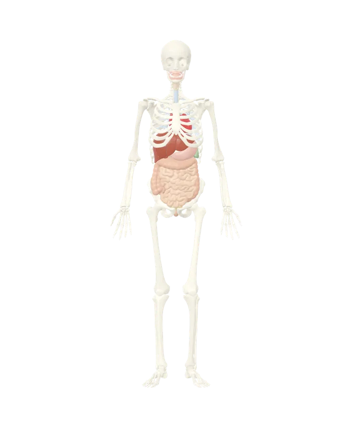
The body opens on the skeleton with the heart, airways and digestive organs in place: 414 of its 1,659 parts.

The skeleton and main organs of the 3D anatomy model, seen from the front

Pick a part

Tap or click any bone, organ or vessel on the model to see its name, what it belongs to and how big it is. Then hide it, show it alone, fade everything else, or pull it out and turn it around.

Body systems

Switch a whole system on or off, or open it to switch its groups and list its parts.

  • Groups in the skeletal system
  • Groups in the muscular system
  • Groups in the cardiovascular system
  • Groups in the nervous system
  • Groups in the respiratory system
  • Groups in the digestive system
  • Groups in the urinary system
  • Groups in the reproductive system
  • Groups in the endocrine system
  • Groups in the lymphatic and immune system
  • Groups in the eye and ear
  • Groups in the skin and hair

The model is one adult male body from the BodyParts3D project, reduced in detail to run on a phone. Real bodies vary, and it is a learning aid, not a clinical reference.

Every one of the 1,659 parts of this body can be tapped, named and moved on its own. Of those, 414 show at the start: the skeleton, the heart, the windpipe and bronchi, the digestive organs, the kidneys and bladder, the spleen, the thymus and four glands. Muscles, blood vessels, the brain, the eyes and the skin sit behind their switches.

What each of the 12 systems holds

The cardiovascular system is the largest in the model at 656 parts, and the lymphatic system the smallest at 3.
System Parts What is in it
Skeletal 281 203 bone parts, 28 teeth, 23 disks, rib and nose cartilages
Muscular 422 Muscles of the head and neck, trunk, arms and legs, with a few tendons
Cardiovascular 656 Heart with chambers and valves, 369 artery parts, 177 vein parts, coronary and lung vessels
Nervous 135 Brain: cortex, deep structures, brainstem, cerebellum and ventricles; nerves of the eye socket
Respiratory 44 Larynx, trachea and the bronchial tree
Digestive 53 Mouth, esophagus, stomach, intestines, liver in eight segments, gallbladder, pancreas
Urinary 6 Two kidneys, two ureters, bladder and urethra
Reproductive 9 Internal male organs
Endocrine 4 Pituitary gland, pineal body and two adrenal glands
Lymphatic and immune 3 Spleen and the two lobes of the thymus
Eye and ear 43 Both eyes layer by layer, eyelids and tear system, outer ears
Skin and hair 3 Skin, hair of the head and eyebrows

Each system loads when its switch goes on, so the first view arrives quickly and the rest follows on request. All twelve systems together come to about 3.3 MB.

Naming a part and taking it out

Tap the left thigh bone. It turns yellow and a card reads Left femur, Skeletal system, Leg and foot, Left side. A short description follows, with the name of the book or agency it came from. Then come what the part belongs to (the left thigh), what kind of structure it is (a long bone) and its size in the model: about 47 by 12 by 6 cm.

Six buttons sit under the card:

  • Zoom to it frames the part.
  • Show only this hides everything else.
  • Fade the rest turns the other parts to glass.
  • Pull it out slides the part clear of the body. A drag then moves it anywhere, and Put it back returns it.
  • Hide it removes the part until Reset.
  • Copy link makes an address that opens the body on that part.

Bones, muscles, organs, the heart, the brain and the eye have written descriptions. Most were drawn from Gray's Anatomy of 1918 and the training modules of the US National Cancer Institute. The smaller arteries and veins show a name, a parent structure and a size only.

With a mouse, pointing at a part shows its name before any click, and a double click zooms straight to it.

Finding the liver and what lies around it

Type liver into the search box. The model holds the liver as eight pieces, and the list shows them: the caudate lobe and segments II to VIII. Pick segment IV. The view closes in on the middle of the liver and the card names it.

Press Fade the rest. The other seven segments, the ribs and the stomach turn to glass, and the segment stands alone in its place under the right side of the rib cage. Press it again, then switch off Skeletal system. Now the whole liver shows with the gallbladder tucked under it, the stomach to its left and the right kidney behind.

The National Cancer Institute places the liver "primarily in the right hypochondriac and epigastric regions of the abdomen, just beneath the diaphragm." It also calls the liver the largest gland in the body. In this model the eight segments together span about 21 cm from side to side and 16 cm from top to bottom. The organs view answers the same kind of question by side and level, and where the liver is covers the organ in full.

Turning, zooming and switching layers

A drag or a swipe turns the body in any direction. Pinch, hold Ctrl and scroll, or press the plus and minus buttons to zoom. Two fingers, or Shift with a drag, slide the view sideways. The round control at the side turns the body when dragged and starts a slow spin when pressed. Front, Side and Back jump to those views, and the home button returns to the start.

Body outline wraps the skin around the scene as glass, which helps place a deep organ against the surface. The pull-apart slider spreads every part away from the center, so close neighbors such as the eight wrist bones and the plates of the skull come apart. Save image downloads the view as a PNG.

Three narrower views start from the same body. The 3D skeleton opens on the bones and adds a naming quiz. The 3D muscles lay 422 muscle parts over the skeleton. The organs view fades everything outside one side or level of the trunk.

Where the model stops

The body is one adult male, about 172 cm tall, from the BodyParts3D project. Some structures a reader might look for are not in the data:

  • Lung surfaces and lobes. The airways are there.
  • The spinal cord and the nerves of the limbs and trunk.
  • Lymph nodes and the thyroid gland.
  • Female organs.
  • The muscles of the face and of chewing.
  • Four trunk muscles: latissimus dorsi, rectus abdominis, internal oblique and transversus abdominis.
  • The ear canal, the eardrum and the three small bones of each ear.

Organs are single smooth shells without inner layers, except the heart, which has its chambers, valves and papillary muscles, and the eye. The model is a learning aid built from one body. Real bodies vary in size and in where their organs sit.

The vessels, brain parts and eye parts by name

495 vessels, brain parts, eye and ear parts. A part that comes as a left and right pair is listed once. Bones are listed with the 3D skeleton, muscles with the 3D muscles and organs with the organs of the body.

Cardiovascular system: coronary vessels (24)
  • Anterior cardiac vein
  • Anterior interventricular vein
  • Circumflex branch of coronary artery
  • Conus artery
  • Conus branch of anterior interventricular branch of coronary artery
  • Coronary sinus
  • Diagonal branch of anterior descending branch of coronary artery
  • First anterior branch of anterior interventricular branch of coronary artery
  • First anterior ventricular branch of coronary artery
  • First posterior ventricular branch of coronary artery
  • First septal branch of posterior interventricular artery
  • Great cardiac vein
  • Marginal branch of coronary artery
  • Marginal vein
  • Middle cardiac vein
  • Posterior interventricular branch of coronary artery
  • Posterior vein of ventricle
  • Second anterior branch of anterior interventricular branch of coronary artery
  • Second septal branch of posterior interventricular artery
  • Septal branch of anterior interventricular artery
  • Small cardiac vein
  • Third anterior branch of anterior interventricular branch of coronary artery
  • Trunk of anterior interventricular branch of coronary artery
  • Trunk of coronary artery
Cardiovascular system: arteries (209)
  • Acromial branch of thoraco-acromial artery
  • Anterior cecal artery
  • Anterior cerebral artery
  • Anterior choroidal artery
  • Anterior circumflex humeral artery
  • Anterior communicating artery
  • Anterior division of renal artery
  • Anterior inferior cerebellar artery
  • Anterior inferior pancreaticoduodenal artery
  • Anterior inferior segmental hepatic artery
  • Anterior interosseous artery
  • Anterior parietal artery
  • Anterior spinal artery
  • Anterior superior pancreaticoduodenal artery
  • Anterior superior segmental hepatic artery
  • Anterior temporal artery
  • Anterior temporal branch of lateral occipital artery
  • Anterior tibial artery
  • Anterior tibial recurrent artery
  • Anterior ulnar recurrent artery
  • Anterolateral central branch of middle cerebral artery
  • Appendicular artery
  • Arch of aorta
  • Arcuate artery
  • Arteria princeps pollicis
  • Arteria radialis indicis
  • Artery of central sulcus
  • Artery of postcentral sulcus
  • Artery of precentral sulcus
  • Ascending aorta
  • Ascending branch of colic artery
  • Ascending branch of inferior branch of ileocolic artery
  • Axillary artery
  • Basilar artery
  • Brachial artery
  • Brachiocephalic artery
  • Branch of anterior choroidal artery to posterior limb of internal capsule
  • Branch of lateral circumflex femoral artery
  • Branch of middle cerebral artery to angular gyrus
  • Bronchial artery
  • Bronchial branch of arch of aorta
  • Callosomarginal artery
  • Caudal pancreatic artery
  • Caudate lobe branch of hepatic artery
  • Celiac trunk
  • Circumflex scapular artery
  • Colic artery
  • Common carotid artery
  • Common hepatic artery
  • Common iliac artery
  • Common interosseous artery
  • Common plantar digital arteries
  • Costocervical trunk
  • Deep brachial artery
  • Deep cervical artery
  • Deep palmar arch
  • Deep plantar artery
  • Deltoid branch of thoraco-acromial artery
  • Descending aorta
  • Descending branch of colic artery
  • Descending branch of lateral circumflex femoral artery
  • Descending genicular artery
  • Distal perforating artery
  • Dorsal carpal branch of radial artery
  • Dorsal carpal branch of ulnar artery
  • Dorsal digital arteries of hand
  • Dorsal digital artery of foot
  • Dorsal metacarpal arteries
  • Dorsal pancreatic artery
  • Dorsal scapular artery
  • Dorsalis pedis artery
  • Esophageal artery
  • Esophageal branches of thoracic aorta
  • External iliac artery
  • Femoral artery
  • Fibular artery
  • First common palmar digital artery
  • First posterior intercostal artery
  • Fourth common palmar digital artery
  • Gastric artery
  • Gastro-epiploic artery
  • Great pancreatic artery
  • Hepatic artery proper
  • Hypothalamic branch of posterior communicating artery
  • Ileal branch of inferior branch of ileocolic artery
  • Ileocolic artery
  • Inferior epigastric artery
  • Inferior lateral genicular artery
  • Inferior medial genicular artery
  • Inferior mesenteric artery
  • Inferior pancreatic artery
  • Inferior pancreaticoduodenal artery
  • Inferior phrenic artery
  • Inferior suprarenal artery
  • Inferior thyroid artery
  • Inferior ulnar collateral artery
  • Insular part of middle cerebral artery
  • Intermediomedial branch of callosomarginal artery
  • Internal carotid artery
  • Internal iliac artery
  • Internal thoracic artery
  • Lateral branch of pontine artery
  • Lateral branch of superior cerebellar artery
  • Lateral circumflex femoral artery
  • Lateral frontobasal artery
  • Lateral inferior segmental hepatic artery
  • Lateral occipital artery
  • Lateral plantar artery
  • Lateral proper palmar digital artery of little finger
  • Lateral proper palmar digital artery of middle finger
  • Lateral proper palmar digital artery of ring finger
  • Lateral superior genicular artery
  • Lateral superior segmental hepatic artery
  • Lateral tarsal artery
  • Lateral thoracic artery
  • Lumbar artery
  • Marginal artery of colon
  • Medial branch of pontine artery
  • Medial branch of superior cerebellar artery
  • Medial frontobasal artery
  • Medial inferior segmental hepatic artery
  • Medial occipital artery
  • Medial plantar artery
  • Medial proper palmar digital artery of index finger
  • Medial proper palmar digital artery of middle finger
  • Medial proper palmar digital artery of ring finger
  • Medial superior genicular artery
  • Medial superior segmental hepatic artery
  • Middle colic artery
  • Middle collateral branch of deep brachial artery
  • Middle genicular artery
  • Middle suprarenal artery
  • Middle temporal branch of lateral occipital artery
  • Middle temporal branch of middle cerebral artery
  • Musculophrenic artery
  • Ophthalmic artery
  • Palmar metacarpal artery
  • Paracentral branch of callosomarginal artery
  • Pectoral branch of thoraco-acromial artery
  • Perforating arteries of thigh
  • Pericallosal artery
  • Plantar arch
  • Plantar metatarsal artery
  • Polar temporal artery
  • Popliteal artery
  • Postcommunicating part of posterior cerebral artery
  • Posterior cecal artery
  • Posterior circumflex humeral artery
  • Posterior communicating artery
  • Posterior division of renal artery
  • Posterior inferior cerebellar artery
  • Posterior inferior pancreaticoduodenal artery
  • Posterior inferior segmental hepatic artery
  • Posterior intercostal arteries
  • Posterior medial choroidal artery
  • Posterior parietal artery
  • Posterior superior pancreaticoduodenal artery
  • Posterior superior segmental hepatic artery
  • Posterior temporal branch of middle cerebral artery
  • Posterior temporal branches of lateral occipital artery
  • Posterior tibial artery
  • Posterior ulnar recurrent artery
  • Posteromedial branch of callosomarginal artery
  • Posteromedial central branch of posterior cerebral artery
  • Precommunicating part of posterior cerebral artery
  • Precuneal branch of pericallosal artery
  • Prefrontal artery
  • Proper plantar digital arteries of foot
  • Radial artery
  • Radial collateral branch of deep brachial artery
  • Radial recurrent artery
  • Recurrent interosseous artery
  • Renal artery
  • Second common palmar digital artery
  • Second posterior intercostal artery
  • Sigmoid artery
  • Sphenoid part of middle cerebral artery
  • Splenial artery
  • Splenic artery
  • Subclavian artery
  • Subcostal artery
  • Subscapular artery
  • Superficial cervical artery
  • Superficial epigastric artery
  • Superficial medial plantar artery
  • Superficial palmar arterial arch
  • Superior cerebellar artery
  • Superior epigastric artery
  • Superior intercostal artery
  • Superior mesenteric artery
  • Superior rectal artery
  • Superior ulnar collateral artery
  • Superior vermian branch of medial branch of superior cerebellar artery
  • Suprascapular artery
  • Temporo-occipital branch of middle cerebral artery
  • Testicular artery
  • Thalamogeniculate artery
  • Thalamoperforating artery
  • Third common palmar digital artery
  • Thoracodorsal artery
  • Thyrocervical trunk
  • Transverse cervical artery
  • Trunk of gastroduodenal artery
  • Trunk of hepatic artery
  • Trunk of inferior terminal branch of middle cerebral artery
  • Trunk of thoraco-acromial artery
  • Ulnar artery
  • Ureteric segment of renal artery
  • Vertebral artery
Cardiovascular system: veins (106)
  • Accessory hemiazygos vein
  • Anterior circumflex humeral vein
  • Anterior inferior segmental branch of portal vein
  • Anterior inferior segmental tributary of middle hepatic vein
  • Anterior intercostal veins
  • Anterior superior segmental branch of portal vein
  • Anterior superior segmental tributary of hepatic vein
  • Anterior tibial vein
  • Ascending lumbar vein
  • Axillary vein
  • Azygos vein
  • Basilic vein
  • Brachiocephalic vein
  • Caudate lobe branch of portal vein
  • Cephalic vein
  • Circumflex scapular vein
  • Colic vein
  • Common iliac vein
  • Common palmar digital vein
  • Deep femoral vein
  • Deep palmar venous arch
  • Dorsal digital veins
  • Dorsal metatarsal veins of foot
  • Dorsal venous arch of foot
  • Dorsal venous network of hand
  • External iliac vein
  • Femoral vein
  • Fibular vein
  • Gastric vein
  • Gastroepiploic vein
  • Genicular vein
  • Great saphenous vein
  • Hemiazygos vein
  • Hepatic portal vein
  • Ileal vein
  • Ileocolic vein
  • Iliolumbar vein
  • Inferior epigastric vein
  • Inferior gluteal vein
  • Inferior mesenteric vein
  • Inferior phrenic vein
  • Inferior vena cava
  • Internal iliac vein
  • Internal jugular vein
  • Internal pudendal vein
  • Internal thoracic vein
  • Lateral circumflex femoral vein
  • Lateral inferior segmental branch of portal vein
  • Lateral inferior segmental tributary of hepatic vein
  • Lateral sacral vein
  • Lateral superior segmental branch of portal vein
  • Lateral superior segmental tributary of hepatic vein
  • Lateral thoracic vein
  • Lumbar vein
  • Medial brachial vein
  • Medial circumflex femoral vein
  • Medial inferior segmental branch of portal vein
  • Medial inferior segmental tributary of middle hepatic vein
  • Medial superior segmental branch of portal vein
  • Medial superior segmental tributary of hepatic vein
  • Median antebrachial vein
  • Median cubital vein
  • Median sacral vein
  • Middle colic vein
  • Musculophrenic vein
  • Obturator vein
  • Palmar metacarpal vein
  • Pancreaticoduodenal vein
  • Perforating veins of foot
  • Perforating veins of thigh
  • Plantar digital veins
  • Plantar metatarsal vein
  • Plantar venous arch of foot
  • Popliteal vein
  • Posterior inferior segmental branch of portal vein
  • Posterior inferior segmental tributary of hepatic vein
  • Posterior superior segmental branch of portal vein
  • Posterior superior segmental tributary of hepatic vein
  • Posterior tibial vein
  • Proper palmar digital vein of index finger
  • Proper palmar digital vein of little finger
  • Proper palmar digital vein of middle finger
  • Proper palmar digital vein of ring finger
  • Radial vein
  • Renal vein
  • Sigmoid vein
  • Small saphenous vein
  • Splenic vein
  • Subclavian vein
  • Subcostal vein
  • Subscapular vein
  • Superficial epigastric vein
  • Superficial palmar venous arch
  • Superior gluteal vein
  • Superior intercostal vein
  • Superior mesenteric vein
  • Superior phrenic vein
  • Superior rectal vein
  • Superior vena cava
  • Suprarenal vein
  • Suprascapular vein
  • Testicular vein
  • Thoracodorsal vein
  • Tributaries of plantar venous arch
  • Trunk of portal vein
  • Ulnar vein
Cardiovascular system: pulmonary arteries (32)
  • Accessory subsuperior branch of posterior basal segmental artery
  • Anterior basal segmental artery
  • Anterior branch of anterior segmental artery
  • Anterior branch of lateral segmental artery
  • Anterior part of apical segmental artery
  • Anterior segmental artery
  • Apical part of apical segmental artery
  • Apical part of posterior segmental artery
  • Apical segmental artery
  • Basal branch of anterior basal segmental artery
  • Inferior branch of medial segmental artery
  • Inferior lingular artery
  • Lateral basal segmental artery
  • Lateral branch of anterior basal segmental artery
  • Lateral branch of lateral basal segmental artery
  • Lateral branch of superior segmental artery
  • Laterobasal branch of posterior basal segmental artery
  • Medial basal segmental artery
  • Medial branch of superior segmental artery
  • Mediobasal branch of posterior basal segmental artery
  • Posterior basal segmental artery
  • Posterior branch of anterior segmental artery
  • Posterior branch of lateral segmental artery
  • Posterior part of posterior segmental artery
  • Posterior segmental artery
  • Pulmonary artery
  • Pulmonary trunk
  • Superior branch of medial segmental artery
  • Superior branch of superior segmental artery
  • Superior lingular artery
  • Superior segmental artery
  • Upper lobar artery
Cardiovascular system: pulmonary veins (21)
  • Anterior basal segmental vein
  • Anterior part of apical segmental vein
  • Anterior part of lateral segmental vein
  • Anterior segmental vein
  • Apical part of apical segmental vein
  • Apical segmental vein
  • Inferior lingular vein
  • Inferior part of anterior segmental vein
  • Inferior part of medial segmental vein
  • Inferior pulmonary vein
  • Lateral basal segmental vein
  • Medial basal segmental vein
  • Posterior basal segmental vein
  • Posterior part of lateral segmental vein
  • Posterior segmental vein
  • Superior lingular vein
  • Superior part of anterior segmental vein
  • Superior part of medial segmental vein
  • Superior pulmonary vein
  • Superior segmental vein
  • Trunk of apical segmental vein
Nervous system: cerebral cortex (18)
  • Angular gyrus
  • Anterior part of superior temporal gyrus
  • Cingulate gyrus
  • Fusiform gyrus
  • Inferior frontal gyrus
  • Inferior temporal gyrus
  • Insula
  • Middle frontal gyrus
  • Middle temporal gyrus
  • Occipital lobe
  • Orbital gyrus
  • Parahippocampal gyrus
  • Postcentral gyrus
  • Posterior part of superior temporal gyrus
  • Precentral gyrus
  • Superior frontal gyrus
  • Superior parietal lobule
  • Supramarginal gyrus
Nervous system: deep brain structures (23)
  • Amygdala
  • Anterior commissure
  • Caudate nucleus
  • Commissure of fornix of forebrain
  • Corpus callosum
  • Fornix of forebrain
  • Globus pallidus
  • Habenula
  • Hippocampus
  • Hypothalamus
  • Internal capsule
  • Lamina terminalis
  • Lateral geniculate body
  • Mammillary body
  • Medial geniculate body
  • Posterior commissure
  • Putamen
  • Septum of telencephalon
  • Stria medullaris of thalamus
  • Stria terminalis
  • Thalamus
  • Tuber cinereum
  • White matter of cerebral hemisphere
Nervous system: brainstem and cerebellum (11)
  • Brachium of inferior colliculus
  • Brachium of superior colliculus
  • Cerebellum
  • Inferior colliculus
  • Interpeduncular fossa
  • Medulla oblongata
  • Midbrain
  • Peduncle of midbrain
  • Pons
  • Superior colliculus
  • Tentorium cerebelli
Nervous system: ventricles (7)
  • Central canal of spinal cord
  • Cerebral aqueduct
  • Choroid plexus of cerebral hemisphere
  • Fourth ventricle
  • Interventricular foramen
  • Lateral ventricle
  • Third ventricle
Nervous system: nerves of the eye socket (19)
  • Anterior ethmoidal nerve
  • Ciliary ganglion
  • Communicating branch of nasociliary nerve with ciliary ganglion
  • Frontal nerve
  • Inferior branch of oculomotor nerve
  • Infratrochlear nerve
  • Lacrimal nerve
  • Long ciliary nerve
  • Nasociliary nerve
  • Ophthalmic nerve
  • Optic chiasm
  • Optic nerve
  • Optic tract
  • Posterior ethmoidal nerve
  • Short ciliary nerve
  • Superior branch of oculomotor nerve
  • Supra-orbital nerve
  • Supratrochlear nerve
  • Trochlear nerve
Eye and ear: eyeball (10)
  • Anterior chamber of eyeball
  • Choroid
  • Cornea
  • Corona ciliaris
  • Iris
  • Lens
  • Optic part of retina
  • Sclera
  • Suspensory ligament of lens
  • Vitreous body
Eye and ear: eyelids and tear system (11)
  • Check ligament of lateral rectus
  • Check ligament of medial rectus
  • Common tendinous ring
  • Lacrimal canaliculus
  • Lacrimal gland
  • Lacrimal lake
  • Lacrimal sac
  • Nasolacrimal duct
  • Tarsal plate of lower eyelid
  • Tarsal plate of upper eyelid
  • Trochlea of superior oblique
Eye and ear: outer ear (1)
  • Outer ears
Skin and hair: skin (1)
  • Skin
Skin and hair: hair (2)
  • Eyebrows
  • Hair of head

More to explore

Questions people ask

How many parts does the 3D human body have?

1,659 named parts in 12 systems. The cardiovascular system is the largest with 656, followed by the muscular system with 422 and the skeletal system with 281. The nervous system has 135, the digestive system 53, the respiratory system 44 and the eye and ear 43.

The four smallest are the reproductive system with 9 parts, the urinary system with 6, the endocrine system with 4 and the lymphatic system with 3. Skin and hair add 3 more.

Can I look at the body layer by layer?

Yes. Each of the 12 systems has its own switch, so the muscles can be laid over the skeleton or taken away again, and the vessels added on top. Inside a system, each group has a switch too: the skull, the spine or the rib cage can be shown alone.

Body outline draws the skin as glass around whatever is showing. Fade the rest turns everything but the chosen part to glass, and the pull-apart slider spreads the parts out from the center of the body.

Is the 3D anatomy model free?

Yes. It runs in the browser with no account and nothing to install. The body data comes from BodyParts3D, a project of Japan's Database Center for Life Science, which shares it under the Creative Commons Attribution 4.0 license.

Is the model a male or a female body?

Male. BodyParts3D describes its data as a whole-body model of an adult human male, and the body stands about 172 cm tall. The reproductive system holds the internal male organs only: the prostate, the seminal vesicles, the deferent ducts, the testes and the epididymides.

There are no female organs and no breast in the data.

Why are there no lungs in the model?

The source data has no lung surfaces. What it has is the airway tree: the trachea, the two main bronchi and 20 segmental bronchial trees, 23 parts in all. Switch on the pulmonary arteries and veins in the cardiovascular system, 61 more parts, and their branching traces where both lungs lie.

How accurate are the names and positions?

The shapes and names come from BodyParts3D, which maps each mesh to a term in a standard anatomy vocabulary. The model here has fewer triangles than the original, about 863,000 against 6.68 million, so fine surface detail is smoothed.

It shows one body. Organs differ in size and position from person to person, so it is a learning aid and not a clinical reference.

4 sources
  1. BodyParts3D, Database Center for Life Science (DBCLS), Japan. Accessed 14 November 2026.
  2. Anatomy & Physiology, SEER Training Modules, National Cancer Institute. Accessed 14 November 2026.
  3. Accessory Organs, SEER Training Modules, National Cancer Institute. Accessed 14 November 2026.
  4. Anatomy of the Human Body, 20th edition (1918), table of contents, Henry Gray, edited by Warren H. Lewis; text hosted by Theodora. Accessed 14 November 2026.