Skip to the main content
3D Body Visualizer

3D brain model: the human brain in 98 named parts

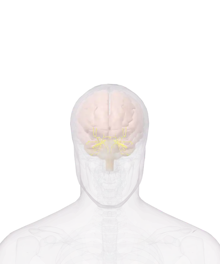
The brain of the 3D anatomy model inside a glass skull, seen from the front

Pick a part

Tap or click a part of the brain for its name, a short description and its size. Hide the cortex to reach the deep parts, or pull a part out and turn it around.

Nervous system: 135 parts

Switch a group on or off, or open its list to pick a part by name.

  • Groups in the nervous system
See the full human anatomy

The brain is that of one adult man, from the BodyParts3D project. The spinal cord and the nerves of the trunk and limbs are not in the model.

The nervous system of the model is the brain: 98 named parts in 4 groups, with 37 nerves of the eye socket as a fifth group. The white matter of each cerebral hemisphere measures 16.3 cm from front to back. The model has no spinal cord and no nerves of the trunk or limbs.

The nervous system of the model in 5 groups

The brain of the model has 98 parts in 4 groups, and 37 nerves of the eye socket make a fifth group.
Group Parts What it holds
Cerebral cortex 36 The folds of the surface, such as the superior frontal gyrus and the cingulate gyrus
Deep brain structures 37 The white matter and the parts that lie under the cortex
Brainstem and cerebellum 16 The midbrain, the pons, the medulla oblongata and the cerebellum
Ventricles 9 The fluid-filled spaces inside the brain
Nerves of the eye socket 37 The nerves that run to the eyes and their muscles

The skull is drawn as glass, so the brain shows in place. Each group has its own switch beside the model. Switch off the cerebral cortex to reach the deep structures and the ventricles.

The 4 divisions of the brain

The brain has 4 divisions, according to the National Cancer Institute: the cerebrum, the diencephalon, the brain stem and the cerebellum.

The cerebrum is the largest of the 4 divisions of the brain and the cerebellum the second largest.
Division What the source says In the model
Cerebrum The largest part, in 2 hemispheres joined by the corpus callosum The cerebral cortex and the white matter of each hemisphere
Diencephalon The thalamus, about 80 percent of it, relays every sense except smell Among the deep brain structures
Brain stem The midbrain, the pons and the medulla oblongata Midbrain, pons and medulla oblongata, each a part
Cerebellum The second largest part, below the occipital lobes One part, behind the brainstem

The medulla oblongata continues into the spinal cord, and all the fibers between the brain and the cord pass through it. Tap the pons in the model, press Pull it out, and the brainstem can be turned around on its own.

The central and the peripheral nervous system

The nervous system has 2 divisions. The central nervous system is the brain and the spinal cord. The peripheral nervous system is the nerves and the ganglia outside them.

The spinal cord runs from the base of the skull to the level of the first lumbar vertebra. It has 31 segments, and each segment gives rise to a pair of spinal nerves. There are 12 pairs of cranial nerves, which leave the brain itself.

Nerve tissue has 2 kinds of cell. Neurons conduct impulses, and neuroglia support them. The National Cancer Institute notes that neuroglia are far more numerous than neurons.

What the model leaves out

The model has no spinal cord, no spinal nerves and no nerves of the trunk, arms or legs. Of the 12 pairs of cranial nerves it shows the ones in the eye socket. The meninges are not drawn, apart from the tentorium cerebelli.

The brain is that of one adult man. The eyes that the nerves of the eye socket serve are in the 3D eye and ear view. The pituitary gland and the pineal body, which sit against the brain, are in the 3D endocrine system, and the bones around the brain are in the 3D skeleton.

Every part of the brain in the model by name

78 brain parts and nerves of the eye socket. A part that comes as a left and right pair is listed once.

Cerebral cortex (18)
  • Angular gyrus
  • Anterior part of superior temporal gyrus
  • Cingulate gyrus
  • Fusiform gyrus
  • Inferior frontal gyrus
  • Inferior temporal gyrus
  • Insula
  • Middle frontal gyrus
  • Middle temporal gyrus
  • Occipital lobe
  • Orbital gyrus
  • Parahippocampal gyrus
  • Postcentral gyrus
  • Posterior part of superior temporal gyrus
  • Precentral gyrus
  • Superior frontal gyrus
  • Superior parietal lobule
  • Supramarginal gyrus
Deep brain structures (23)
  • Amygdala
  • Anterior commissure
  • Caudate nucleus
  • Commissure of fornix of forebrain
  • Corpus callosum
  • Fornix of forebrain
  • Globus pallidus
  • Habenula
  • Hippocampus
  • Hypothalamus
  • Internal capsule
  • Lamina terminalis
  • Lateral geniculate body
  • Mammillary body
  • Medial geniculate body
  • Posterior commissure
  • Putamen
  • Septum of telencephalon
  • Stria medullaris of thalamus
  • Stria terminalis
  • Thalamus
  • Tuber cinereum
  • White matter of cerebral hemisphere
Brainstem and cerebellum (11)
  • Brachium of inferior colliculus
  • Brachium of superior colliculus
  • Cerebellum
  • Inferior colliculus
  • Interpeduncular fossa
  • Medulla oblongata
  • Midbrain
  • Peduncle of midbrain
  • Pons
  • Superior colliculus
  • Tentorium cerebelli
Ventricles (7)
  • Central canal of spinal cord
  • Cerebral aqueduct
  • Choroid plexus of cerebral hemisphere
  • Fourth ventricle
  • Interventricular foramen
  • Lateral ventricle
  • Third ventricle
Nerves of the eye socket (19)
  • Anterior ethmoidal nerve
  • Ciliary ganglion
  • Communicating branch of nasociliary nerve with ciliary ganglion
  • Frontal nerve
  • Inferior branch of oculomotor nerve
  • Infratrochlear nerve
  • Lacrimal nerve
  • Long ciliary nerve
  • Nasociliary nerve
  • Ophthalmic nerve
  • Optic chiasm
  • Optic nerve
  • Optic tract
  • Posterior ethmoidal nerve
  • Short ciliary nerve
  • Superior branch of oculomotor nerve
  • Supra-orbital nerve
  • Supratrochlear nerve
  • Trochlear nerve

More to explore

Questions people ask

What are the main parts of the brain?

The brain has 4 main divisions: the cerebrum, the diencephalon, the brain stem and the cerebellum. The National Cancer Institute calls the cerebrum the largest part of the brain and the cerebellum the second largest.

How many cranial nerves and spinal nerves are there?

There are 12 pairs of cranial nerves and 31 pairs of spinal nerves. Of the cranial nerves, 3 pairs are sensory, 5 are mainly motor and 4 are mixed. The spinal nerves are 8 cervical, 12 thoracic, 5 lumbar, 5 sacral and 1 coccygeal pair.

Is the spinal cord in the 3D model?

No. The model has the brain and 37 nerves of the eye socket, and no spinal cord and no nerves of the trunk or limbs. The National Cancer Institute places the spinal cord in the vertebral canal, from the opening at the base of the skull to the first lumbar vertebra.

What does the thalamus do?

The thalamus relays sensory impulses to the cerebral cortex, for every sense except smell, according to the National Cancer Institute. The thalamus makes up about 80 percent of the diencephalon. In the model it sits among the deep brain structures, under the cortex.

What covers the brain?

Three membranes called meninges cover the brain and the spinal cord. The dura mater is the tough outer layer, the arachnoid is the middle layer, and the pia mater is the inner layer bound to the surface of the brain. The model does not draw the meninges, apart from the tentorium cerebelli.

6 sources
  1. Introduction to the Nervous System, SEER Training Modules, National Cancer Institute. Accessed 14 November 2026.
  2. Organization of the Nervous System, SEER Training Modules, National Cancer Institute. Accessed 14 November 2026.
  3. The Central Nervous System, SEER Training Modules, National Cancer Institute. Accessed 14 November 2026.
  4. The Peripheral Nervous System, SEER Training Modules, National Cancer Institute. Accessed 14 November 2026.
  5. Nerve Tissue, SEER Training Modules, National Cancer Institute. Accessed 14 November 2026.
  6. BodyParts3D, Database Center for Life Science (DBCLS), Japan. Accessed 14 November 2026.