Skip to the main content
3D Body Visualizer

Female reproductive anatomy in 3D: uterus, ovaries and tubes, labeled

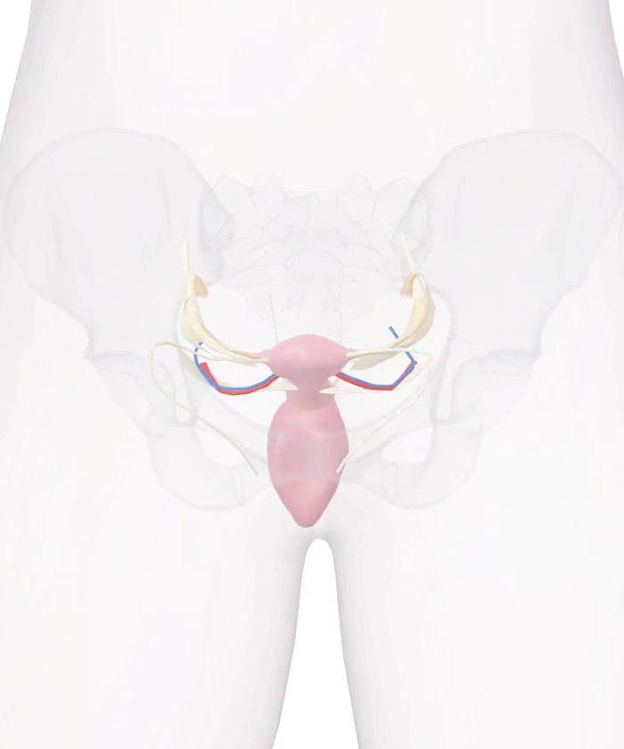
The uterus, ovaries and uterine tubes of the 3D model inside a glass female pelvis, seen from the front
FemaleMale

Pick a part

Tap or click the uterus, an ovary, a tube or a ligament to see its name and its size. Then hide it, show it alone, or pull it out of the pelvis and turn it around.

Female reproductive system: 41 parts

Switch a group on or off, or open its list to pick a part by name.

  • Groups in the female reproductive system
See the full human anatomy

The organs are those of one adult woman, from the Human Reference Atlas of the National Institutes of Health. She is a different person from the male body of the other views. Organs differ in size and position from person to person.

The uterus of this model is 5.2 cm across, and each of its 2 ovaries is 2.4 cm long: 41 parts of one woman's pelvic organs, each with its name.

The female reproductive anatomy of this model has 41 parts: the uterus and its cervix, 2 ovaries, 2 uterine tubes, the vagina, 16 ligaments and folds, and 4 uterine vessels. The bones of the female pelvis are drawn as glass around them, inside the outline of the body. The organs are those of one adult woman, 166.6 cm tall.

The 41 parts of the model

The 41 parts of the model fall into 8 groups.
Group Parts What it holds
Uterus and cervix 7 Body, fundus, horns, lower segment, cervix and its 2 openings
Walls of the uterus 2 Anterior and posterior wall
Ovaries 2 Left and right ovary
Uterine tubes 8 Isthmus, ampulla, infundibulum and fimbriae on each side
Vagina 2 Vagina and its junction with the cervix
Ligaments 14 7 pairs, from the round ligaments to the mesovarium
Broad ligament and pouch 2 Broad ligament and uterovesical pouch
Uterine vessels 4 Uterine artery and vein on each side

The walls of the uterus and the broad ligament start switched off, because they cover the parts under them. Switch them on in the list of groups. Tap any part and press "Fade the rest" to see it alone inside the pelvis.

What each organ does

The ovaries make the egg cells. The National Cancer Institute counts about 700,000 egg cells in the two ovaries at birth and about 400,000 at puberty, and none are made after birth. From puberty, one follicle usually ripens each month.

The uterine tubes carry the egg to the uterus. The egg can be fertilized for only 24 to 48 hours, and that usually happens in the tube. The journey down the tube takes about 7 days.

The uterus is a hollow organ with a thick wall of muscle. Gray's Anatomy places it deep in the pelvis, between the bladder in front and the rectum behind. The lining of the uterus has 2 layers: one is shed at menstruation, and the deeper one rebuilds it.

The cervix is the lower, narrow part of the uterus. It projects into the vagina, which leads to the outside of the body.

Sizes of the organs in the adult, from the two sources.
Organ Size in the adult Source
Ovary About 3.5 cm long, 2 cm wide, 1 cm thick National Cancer Institute
Ovary About 4 cm long, 2 cm wide, 8 mm thick; 2 to 3.5 grams Gray's Anatomy
Uterine tube About 10 cm long Gray's Anatomy
Uterus About 7.5 cm long, 5 cm broad, nearly 2.5 cm thick; 30 to 40 grams Gray's Anatomy
Vagina 6 to 7.5 cm along its front wall, 9 cm along its back wall Gray's Anatomy
Vagina About 10 cm long National Cancer Institute

The ligaments that hold the organs

The uterus is not fixed to bone. Folds of the lining of the abdomen and cords of fibrous tissue hold it and the ovaries in place.

Six ligaments and folds hold the uterus, the tubes and the ovaries in place.
Ligament What it joins
Broad ligament The sides of the uterus to the side walls of the pelvis; the tubes and ovaries lie between its 2 layers
Round ligament The upper corner of the uterus, through the groin, to the labium majus; 10 to 12 cm long
Ligament of the ovary The lower end of the ovary to the upper corner of the uterus
Suspensory ligament of the ovary The upper end of the ovary to the side wall of the pelvis; it carries the vessels of the ovary
Uterosacral ligament The cervix to the front of the sacrum
Cardinal ligament The side of the cervix and the top of the vagina to the side wall of the pelvis

The mesosalpinx is the part of the broad ligament between the tube and the ovary. The mesovarium is a short fold that joins the ovary to the back of the broad ligament. The uterovesical pouch is the pocket between the front of the uterus and the bladder.

The uterine artery on each side is a branch of the internal iliac artery. It meets the artery of the ovary, which comes from the aorta, and the two supply the uterus.

The female pelvis

The pelvis of the model is 31.7 cm wide and 21.1 cm high. It has 8 parts: the sacrum, the coccyx, and the ilium, ischium and pubis of each side. Gray's Anatomy describes the female pelvis as more delicate than the male, with a shallower and wider cavity, a larger and more nearly circular upper opening, and a wider and more rounded pubic arch. The differences show as early as the fourth month before birth.

What the model leaves out

The model has no external organs, no breasts and no bladder or rectum. It does not draw the inside of the uterus or the 3 layers of its wall. The source names the 2 round ligaments with their sides exchanged, so each one is named here by the side it sits on. The organs are those of one adult woman, and their size and position differ from person to person.

The male organs are in the 3D male reproductive system. The bladder that lies in front of the uterus is in the 3D urinary system, and the pituitary gland that controls the ovaries is in the 3D endocrine system. The male pelvis is part of the 3D skeleton.

Every part of the female reproductive organs in the model by name

27 parts of the uterus, ovaries, tubes, vagina, ligaments and vessels. A part that comes as a left and right pair is listed once.

Uterus and cervix (7)
  • Body of uterus
  • Cervix of uterus
  • External os of cervix
  • Fundus of uterus
  • Horns of uterus
  • Internal os of cervix
  • Lower uterine segment
Walls of the uterus (2)
  • Anterior wall of uterus
  • Posterior wall of uterus
Ovaries (1)
  • Ovary
Uterine tubes (4)
  • Ampulla of uterine tube
  • Fimbriae of uterine tube
  • Infundibulum of uterine tube
  • Isthmus of uterine tube
Vagina (2)
  • Cervicovaginal junction
  • Vagina
Ligaments (7)
  • Cardinal ligament of uterus
  • Ligament of ovary
  • Mesosalpinx
  • Mesovarium
  • Round ligament of uterus
  • Suspensory ligament of ovary
  • Uterosacral ligament
Broad ligament and pouch (2)
  • Broad ligament of uterus
  • Uterovesical pouch
Uterine vessels (2)
  • Uterine artery
  • Uterine vein

The uterus is a hollow organ with a thick wall of muscle, deep in the pelvis between the bladder in front and the rectum behind. In the adult it is about 7.5 cm long, 5 cm broad and nearly 2.5 cm thick, and weighs 30 to 40 grams. A slight narrowing about halfway down divides it into the body above and the cervix below.

The fundus is the rounded top of the uterus, the part of the body that lies above the openings of the two uterine tubes.

The cervix is the lower, narrow segment of the uterus, somewhat conical in shape. It projects through the front wall of the vagina. Its internal opening leads up into the body of the uterus, and its external opening leads down into the vagina.

The ovaries make the egg cells. Each is about 3.5 cm long, 2 cm wide and 1 cm thick, about the size and shape of an almond, and sits in a shallow hollow of the side wall of the pelvis beside the uterus. The two ovaries hold about 700,000 egg cells at birth and about 400,000 at puberty.

Each uterine tube is about 10 cm long and carries the egg from the ovary to the uterus. Its narrow inner third is the isthmus, its wider middle part is the ampulla, which curves over the ovary, and its funnel-shaped end is the infundibulum, edged with fimbriae. One fimbria is attached to the ovary.

The vagina extends from the outside of the body to the uterus, behind the bladder and in front of the rectum. It measures 6 to 7.5 cm along its front wall and 9 cm along its back wall, and it surrounds the lower part of the cervix.

The round ligaments are two cords, each between 10 and 12 cm long. Each starts at the upper corner of the uterus, passes through the inguinal canal in the groin and ends in the labium majus.

The uterosacral ligaments are folds of fibrous tissue and muscle that run back from the cervix to the front of the sacrum. The cardinal ligaments are attached to the side of the cervix and to the top of the vagina.

The ligament of the ovary is a rounded cord from the lower end of the ovary to the upper corner of the uterus, just behind the uterine tube. The suspensory ligament is a fold from the upper end of the ovary to the side wall of the pelvis, and it carries the vessels of the ovary.

The two broad ligaments pass from the sides of the uterus to the side walls of the pelvis. Between their two layers lie the uterine tube, the round ligament, the ovary and its ligament, and vessels. The mesosalpinx is the part between the tube and the level of the ovary, and the mesovarium is a short fold that joins the ovary to the back of the broad ligament.

The uterovesical pouch is the pocket where the lining of the abdomen passes from the front of the uterus onto the bladder.

The uterine artery on each side is a branch of the internal iliac artery. It meets the artery of the ovary, which comes from the aorta, and the two supply the uterus along a winding course. The veins follow the arteries.

More to explore

Questions people ask

What are the parts of the female reproductive system?

The internal organs are the 2 ovaries, the 2 uterine tubes, the uterus and the vagina. The ovaries make the egg cells, the tubes carry them to the uterus, and the vagina leads from the uterus to the outside of the body.

The model draws those organs in 21 parts, with 16 ligaments and folds that hold them and 4 uterine vessels.

How big is the uterus?

Gray's Anatomy gives the adult uterus as about 7.5 cm long, 5 cm broad and nearly 2.5 cm thick, with a weight of 30 to 40 grams. The National Cancer Institute describes it as about the size and shape of a pear.

The uterus of this model is 5.2 cm across at its widest.

Where are the ovaries, and how big are they?

Each ovary sits in a shallow hollow of the side wall of the pelvis, beside the uterus. The National Cancer Institute gives its size as about 3.5 cm long, 2 cm wide and 1 cm thick, about the size and shape of an almond.

Each ovary of the model is 2.4 cm long.

How long are the uterine tubes?

Each uterine tube is about 10 cm long, according to Gray's Anatomy. It has 3 parts: a narrow isthmus next to the uterus, a wider ampulla that curves over the ovary, and a funnel called the infundibulum, edged with finger-like fimbriae.

How does the female pelvis differ from the male pelvis?

Gray's Anatomy lists the differences. The bones of the female pelvis are more delicate, its cavity is shallower and wider, and its upper opening is larger and more nearly circular. The pubic arch is wider and more rounded, where the male pelvis has an angle.

The pelvis of this model is 31.7 cm wide.

Is this the same body as the rest of the 3D anatomy?

No. The other anatomy views are one adult male body. The female organs come from a second body, an adult woman 166.6 cm tall, so they are shown with her own pelvis and her own outline and are never mixed into the male body.

8 sources
  1. Ovaries, SEER Training Modules, National Cancer Institute. Accessed 15 November 2026.
  2. Genital Tract, SEER Training Modules, National Cancer Institute. Accessed 15 November 2026.
  3. The Uterus, Gray's Anatomy of the Human Body (1918), online edition at Theodora.com. Accessed 15 November 2026.
  4. The Uterine Tube, Gray's Anatomy of the Human Body (1918), online edition at Theodora.com. Accessed 15 November 2026.
  5. The Ovaries, Gray's Anatomy of the Human Body (1918), online edition at Theodora.com. Accessed 15 November 2026.
  6. The Vagina, Gray's Anatomy of the Human Body (1918), online edition at Theodora.com. Accessed 15 November 2026.
  7. The Pelvis, Gray's Anatomy of the Human Body (1918), online edition at Theodora.com. Accessed 15 November 2026.
  8. CCF 3D Reference Object Library, Human Reference Atlas, National Institutes of Health. Accessed 15 November 2026.