Skip to the main content
Body Visualizer 3D

Organs of the body in 3D: which organ sits where

Choose a side and a level, and the organs that sit there stay solid while the rest of the trunk turns to glass.

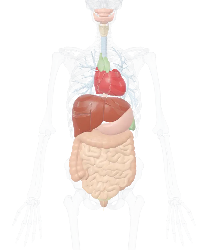
The organs of the 3D anatomy model, seen from the front

What sits where

Pick a side, a level or both. The organs there stay solid and the rest fade.

Pick a part

Tap or click an organ for its name, a short description and its size. Hide it to see what lies behind, or pull it out and turn it around.

Body systems

Switch a whole system on or off, or open it to switch its groups and list its parts.

  • Groups in the skeletal system
  • Groups in the muscular system
  • Groups in the cardiovascular system
  • Groups in the nervous system
  • Groups in the respiratory system
  • Groups in the digestive system
  • Groups in the urinary system
  • Groups in the reproductive system
  • Groups in the endocrine system
  • Groups in the lymphatic and immune system
  • Groups in the eye and ear
  • Groups in the skin and hair

The organs are those of one adult man, from the BodyParts3D project, and the lungs are not among them. Where an organ sits and how large it is differ from person to person.

The liver sits mainly on the right, and the stomach and spleen in the upper left. The heart lies in the chest with two-thirds of its mass left of the midline. Those are the National Cancer Institute's descriptions, and the body on screen shows each of them. Its organs are solid, its skeleton is glass, and two rows of buttons pick out one side or one level at a time.

Organs by side and level in this body

For this body the finder lists 11 organs in the right upper abdomen and 11 in the left upper abdomen.
Level Right side Middle Left side
Chest Bronchi Heart, trachea, esophagus, thymus, bronchi Heart, bronchi
Upper abdomen Liver, gallbladder, bile ducts, duodenum, pancreas, kidney, adrenal gland, ascending colon, transverse colon, jejunum, ureter Liver, stomach, pancreas, duodenum, bile ducts, transverse colon Stomach, spleen, pancreas, liver, bile ducts, kidney, adrenal gland, descending colon, transverse colon, jejunum, ureter
Lower abdomen and pelvis Appendix, ascending colon, ileocecal junction, ileum, ureter Bladder, urethra, rectum, ileum, jejunum, appendix Descending colon, ileum, jejunum, ureter

Every cell is what the finder answers for that pair of buttons. A fourth level, Head and neck, holds the tongue, lips, gums, salivary glands, larynx, pituitary gland and pineal body, all on or near the midline.

Left and right are the body's own. From the front, the body's left is on the viewer's right.

How the finder decides

Each organ is a surface made of thousands of points. The finder counts how many of an organ's points fall in the chosen region and names the organ when the share reaches 18 percent. The middle is a band 3 cm either side of the midline, so the right and left sides begin 3 cm out.

Levels work in two ways. The chest means the organs of the chest cavity: the heart, the airways, the esophagus and the thymus. Height alone cannot separate chest from abdomen, since the dome of the liver rises higher than the base of the heart. The abdomen is split by height, at the disk under the third lumbar vertebra, 105 cm from the floor in this body.

The rule is simple and it runs on one body. An organ near a boundary can fall either way, which is why the appendix shows under both Right side and Middle here.

What lies under the right ribs

Press Right side, then Upper abdomen. Eleven organs stay solid and a sentence names them: the adrenal gland, ascending colon, bile ducts, duodenum, gallbladder, jejunum, kidney, liver, pancreas, transverse colon and ureter.

The gallbladder hides behind the liver from the front, so type gallbladder into the search box and pick it. Its card reads Gallbladder, Digestive system, Liver and bile ducts, with a description and a size of about 5 by 4 by 4 cm. Press Pull it out and the green sac slides clear of the liver, where it can be seen from every side.

Now press Middle as well. The stomach joins the list, since the finder adds regions together when more than one button is on. Reset clears every choice.

Six organs, where they sit and how big they are here

The stomach of this model measures 13.4 cm from side to side and the gallbladder 4.8 cm from front to back.
Organ Where it sits Largest dimension in this model
Liver Mainly the right upper abdomen, just beneath the diaphragm 20.9 cm side to side, in eight segments
Stomach Upper left quadrant of the abdomen 13.4 cm side to side
Spleen Upper left abdomen, beneath the diaphragm and behind the stomach 9.5 cm front to back
Kidneys Either side of the spine, twelfth thoracic to third lumbar vertebra 10.6 cm and 10.4 cm tall
Gallbladder Attached to the visceral surface of the liver 4.8 cm front to back
Heart In the chest, two-thirds of its mass left of the midline Ventricles 10.2 cm side to side

The positions are the National Cancer Institute's, from its SEER training modules. The sizes belong to this one body.

Switching organs and systems on and off

The body opens with 133 organ parts in place: the digestive organs, the airways, the kidneys and bladder, the heart with its valves, the spleen, the thymus and four glands. Under Body systems, each can be switched off to see what lies behind it. Switch off the small intestine, for example, and whatever its coils were hiding comes into view.

The skeleton stays as glass for bearings and turns solid with its own switch. Arteries and veins, muscles and the internal male organs can be added the same way. Body outline draws the skin around the whole scene, which shows how deep each organ lies.

What this body cannot show

There are no lung surfaces, only the airways inside them. There is no thyroid gland, no lymph nodes and no female organs. The model has no separate cecum or sigmoid colon. Organs are smooth outer shells without inner layers, apart from the heart.

One adult man's organs are shown. Size and position differ from person to person. The lists are a guide for learning, and they cannot explain a pain. A doctor can examine the real thing.

The organs on the right and left side are set out organ by organ in their own guide, and the abdominal quadrants divide the same ground into four. The full 3D anatomy view opens the same body with all 12 systems, and the 3D skeleton and 3D muscles show what surrounds the organs.

Every organ part of the model by name

110 organ parts. A part that comes as a left and right pair is listed once.

Cardiovascular system: heart (18)
  • Anterior cusp of aortic valve
  • Anterior cusp of pulmonary valve
  • Anterior leaflet of mitral valve
  • Anterior leaflet of tricuspid valve
  • Anterior papillary muscle of ventricle
  • Anterolateral head of lateral papillary muscle of ventricle
  • Cavity of atrium
  • Cavity of ventricle
  • Lateral papillary muscle of ventricle
  • Posterior cusp of aortic valve
  • Posterior cusp of pulmonary valve
  • Posterior leaflet of mitral valve
  • Posterior leaflet of tricuspid valve
  • Posterior papillary muscle of ventricle
  • Septal leaflet of tricuspid valve
  • Septal papillary muscle of ventricle
  • Wall of atrium
  • Walls of the ventricles
Respiratory system: larynx (14)
  • Arytenoid cartilage
  • Conus elasticus
  • Corniculate cartilage
  • Cricoid cartilage
  • Cuneiform cartilage
  • Epiglottis
  • Hyo-epiglottic ligament
  • Lateral thyrohyoid ligament
  • Median cricothyroid ligament
  • Median thyrohyoid ligament
  • Thyro-epiglottic ligament
  • Thyrohyoid membrane
  • Thyroid cartilage
  • Vocal ligament
Respiratory system: trachea and bronchi (14)
  • Anterior basal segmental bronchial tree
  • Anterior segmental bronchial tree
  • Apical segmental bronchial tree
  • Inferior lingular bronchial tree
  • Lateral basal segmental bronchial tree
  • Lateral segmental bronchial tree
  • Main bronchus
  • Medial basal segmental bronchial tree
  • Medial segmental bronchial tree
  • Posterior basal segmental bronchial tree
  • Posterior segmental bronchial tree
  • Superior lingular bronchial tree
  • Superior segmental bronchial tree
  • Trachea
Digestive system: mouth and throat (6)
  • Gingiva of lower jaw
  • Gingiva of upper jaw
  • Lips
  • Sublingual gland
  • Submandibular gland
  • Tongue
Digestive system: esophagus and stomach (2)
  • Esophagus
  • Stomach
Digestive system: small intestine (8)
  • Distal part of ileum
  • Distal part of jejunum
  • Duodenum
  • Mesentery of small intestine
  • Middle part of ileum
  • Middle part of jejunum
  • Proximal part of ileum
  • Proximal part of jejunum
Digestive system: large intestine (11)
  • Appendix
  • Ascending colon
  • Descending colon
  • Ileocecal junction
  • Mesoappendix
  • Rectum
  • Taenia libera
  • Taenia mesocolica
  • Taenia omentalis
  • Transverse colon
  • Transverse mesocolon
Digestive system: liver and bile ducts (21)
  • Anterior inferior tributary of hepatic biliary tree
  • Anterior superior tributary of hepatic biliary tree
  • Caudate lobe of liver
  • Common hepatic duct
  • Cystic duct
  • Duct of caudate lobe of liver
  • Gallbladder
  • Hepatic duct
  • Lateral inferior tributary of hepatic biliary tree
  • Lateral superior tributary of hepatic biliary tree
  • Liver segment II
  • Liver segment III
  • Liver segment IV
  • Liver segment V
  • Liver segment VI
  • Liver segment VII
  • Liver segment VIII
  • Medial inferior tributary of hepatic biliary tree
  • Medial superior tributary of hepatic biliary tree
  • Posterior inferior tributary of hepatic biliary tree
  • Posterior superior tributary of hepatic biliary tree
Digestive system: pancreas (2)
  • Pancreas
  • Pancreatic ducts
Urinary system: kidneys (1)
  • Kidney
Urinary system: ureters, bladder and urethra (3)
  • Ureter
  • Urethra
  • Urinary bladder
Reproductive system: male internal organs (5)
  • Deferent duct
  • Epididymis
  • Prostate
  • Seminal vesicle
  • Testis
Endocrine system: glands (3)
  • Adrenal gland
  • Pineal body
  • Pituitary gland
Lymphatic and immune system: spleen and thymus (2)
  • Lobe of thymus
  • Spleen

More to explore

Questions people ask

What organs are on the right side of the body?

In this model the right side holds most of the liver, the gallbladder, the right kidney and adrenal gland, the duodenum, part of the pancreas, the ascending colon and the appendix. Bronchi of the right lung run through the right side of the chest.

The National Cancer Institute places the liver primarily in the right upper part of the abdomen, just beneath the diaphragm.

What organs are on the left side of the body?

The spleen, most of the stomach, the left kidney and adrenal gland, part of the pancreas, the descending colon and much of the jejunum. The heart also counts for the left: the National Cancer Institute puts two-thirds of its mass to the left of the midline.

The left lobe of the liver crosses the midline too, so the liver appears under both sides.

Is the heart on the left side of the chest?

Mostly. The National Cancer Institute describes the heart as shaped and sized roughly like a closed fist, with two-thirds of its mass to the left of the midline. The finder agrees for this body: it lists the heart under Middle and under Left side, and not under Right side.

Where are the kidneys?

At the back of the upper abdomen, one on each side of the spine. The National Cancer Institute places them between the twelfth thoracic and third lumbar vertebrae, and says the right kidney usually sits slightly lower because the liver pushes it down.

The model shows that: its right kidney lies about 2 cm lower than its left. Turn the body to the back view to see both against the lowest ribs.

Why does one organ show up on both sides?

Some organs cross the midline. The liver's left lobe reaches into the left side, the pancreas lies across the spine, and the transverse colon runs from one flank to the other.

The finder names an organ for a region when at least 18 percent of its surface lies there, so an organ that spans two regions is listed in both.

Which side is left when I look at the model?

Left and right always mean the body's own. Seen from the front, the body's left is on your right, the same as when facing another person. Each part's card states its side, such as Left kidney, Urinary system, Left side.

Where are the lungs?

The body's data has no lung surfaces, so the finder cannot list them. The airways are there: the trachea, the two main bronchi and the bronchial trees that branch through both lungs. They show where each lung lies, to the right and left of the heart.

Where is the spleen?

In the upper left of the abdomen. The National Cancer Institute places the spleen in the upper left abdominal cavity, just beneath the diaphragm and behind the stomach. The finder lists it under Left side and Upper abdomen only. In this model the spleen measures 9.5 cm from front to back.

Where is the stomach?

In the upper left of the abdomen. The National Cancer Institute puts the stomach in the upper left quadrant, where it receives food from the esophagus. On this body a little over half of the stomach lies left of the middle band and the rest sits on the midline, so the finder lists it under Middle and Left side.

Which side is the appendix on?

The right. The National Institute of Diabetes and Digestive and Kidney Diseases describes the appendix as a finger-like pouch attached to the large intestine, in the lower right area of the abdomen. The finder lists it under Lower abdomen and pelvis, on the Right side, with part of it reaching the middle band on this body.

7 sources
  1. Accessory Organs, SEER Training Modules, National Cancer Institute. Accessed 14 November 2026.
  2. Stomach, SEER Training Modules, National Cancer Institute. Accessed 14 November 2026.
  3. Spleen, SEER Training Modules, National Cancer Institute. Accessed 14 November 2026.
  4. Kidneys, SEER Training Modules, National Cancer Institute. Accessed 14 November 2026.
  5. Structure of the Heart, SEER Training Modules, National Cancer Institute. Accessed 14 November 2026.
  6. Definition & Facts for Appendicitis, National Institute of Diabetes and Digestive and Kidney Diseases. Accessed 14 November 2026.
  7. BodyParts3D, Database Center for Life Science (DBCLS), Japan. Accessed 14 November 2026.